top of page

The Future of 3D Tissue Maps
May 11, 2025
In-person at Harvard Medical School
About
Recent technological advances enable the generation of high-resolution 3D tissue maps down to the single-cell level, revealing new insights into tissue architectures and their biological significance in healthy and diseased organs.
Visualization and user interaction with 3D tissue maps is a critical frontier because it enables deeper insights into the intricate architecture and function of biological tissues. High-resolution 3D tissue maps generated through advanced imaging and spatial omics technologies capture cellular organization, spatial relationships, and molecular distributions with unprecedented detail. However, these datasets are vast, complex, and multidimensional, making it challenging to extract meaningful biological insights without effective tools for exploration and analysis. Interactive visualizations provide an intuitive way to navigate and interrogate these data, uncovering patterns and relationships that are difficult to discern through computational methods alone. By empowering scientists to interact directly with these maps, we can enhance hypothesis generation, drive discoveries in disease mechanisms, and advance precision medicine approaches that depend on spatially resolved data.
This event brings together leading experts in 3D spatial biology to address these challenges and explore opportunities for innovation in data generation, computational analysis, and interactive visualization of spatial omics and imaging data. It will be a unique opportunity to network with biologists, software developers, data visualization specialists, UI/UX designers, and other professionals across career stages. The event will provide opportunities to learn about the latest progress in the field, exchange insights, discuss challenges and solutions with your peers, and foster collaborations to shape the future of 3D spatial biology.
This free, one-day in-person event is part of the virtual VUES on Spatial Biology Seminar Series supported by the NIH Human BioMolecular Atlas Program and hosted by the HIDIVE Lab in the Department of Biomedical Informatics at Harvard Medical School.
Speakers

Ashley Kiemen, PhD
Department of Pathology, Johns Hopkins University
Dr. Ashley Kiemen received a bachelor’s degree in chemical engineering from the University of Michigan, a master’s degree in philosophy from the London School of Economics, and a PhD in chemical & biomolecular engineering from Johns Hopkins University. She is an Assistant Professor of Pathology at the Johns Hopkins School of Medicine. Ashley’s work focuses on using deep learning and digital pathology to study the early development and invasion patterns of pancreatic cancer in three dimensions. Her group developed CODA , a technique to combine serial histological sectioning with spatial transcriptomics, genomics, and proteomics to deeply profile cm-sized tissues in 3D.

Clarence Yapp, DPhil
Harvard Medical School
Clarence Yapp is the Director for Microscopy and Computer Vision at the Laboratory of Systems Pharmacology where he advises on the collection and analysis of highly multiplex image data (CYCIF) for studying disease. Recently, he has extended CyCIF to 3D imaging methods with strong collaboration with the Danuser lab (UTSW) and Gehlenborg lab (HIDIVE@HMS). Clarence comes from a robotics and biomedical engineering background, and used his experiences to develop computer vision tools and microscopy-based assays for studying diseases. For his PhD at the University of Oxford, he pioneered an optical technique that introduced non-permeable molecules into diseased cells using a multiphoton laser. This technique was later adopted to assist in drug discovery against novel epigenetic targets such as bromodomains and demethylases. He moved to Boston in 2016 where he had additional roles at the Image and Data Analysis Core (IDAC), and the Institute of Chemistry and Cell Biology (ICCB-L) where he advised on assay design for the entire HMS and the nearby 3 research hospitals. Clarence frequently promotes open-source tools and is a lead developer in the popular MCMICRO end-to-end image analysis pipeline.

Eric Moerth, PhD
Harvard Medical School - Department of Biomedical Informatics (DBMI)
Eric Moerth is a Post Doctoral Specialist Research Fellow in the Department of Biomedical Informatics at Harvard Medical School (DBMI) and part of the HIDIVE Lab. He received his PhD from the University of Bergen in Norway. During his PhD study, Eric Moerth conducted research in multimodal medical visualization. His focus was the research of new and innovative ways to visualize and explore medical data. Now Eric is researching the visualization of large-scale 3D Tissue Images using novel rendering techniques. Furthermore, his research enables the efficient analysis of highly multiplexed tissue images, and he also investigates how novel output devices like virtual and augmented reality devices can help investigators make better sense of their data.

Gloria Pryhuber, MD
University of Rochester Medical Center
I am a physician-scientist with more than 30 years as an principal investigator in translational and basic science of lung injury and neonatal immunology, I foster active collaborations of investigators and clinicians in neonatology, pulmonology, infectious disease, obstetrics/perinatology, immunology, microbiology, pathology, molecular biology and genomics, aging effects (from neonate to elderly adult) on function and lung disease. My broad goal is to promote collaborative research to reduce respiratory mortality and morbidity in children and adults. In addition to managing several observational studies in neonatology, including the NIH multicenter Environmental Influences on Child Health Outcomes (ECHO) program, I maintain a basic science laboratory, serve as PI for the Lung Development Molecular Atlas Program (LungMAP) Human Tissue Core (HTC) BioRepository for INvestigation of Diseases of the Lung (BRINDL) now in our 11th year, and as Contact PI for the lung component of the NIH Human Biomolecular Atlas Program (HuBMAP - Lung). As the LungMAP HTC, in partnership with the UNOS national transplant network, provide an extensive and deeply annotated repository of transplant-quality, research-authorized tissues including trachea and lung tissue, peripheral blood mononuclear cells (PBMC), lymph node, spleen and thymus from over 470 donors, newborn to elderly. As such, we provide multiple research centers with opportunities to explore the developing human respiratory tract and immune system in an unusually holistic manner. See LungMAP.net, HuBMAP and LGEA web portals for the work of my laboratory and our collaborators using these samples to generate and make publicly available reference sc/snRNAseq and spatial transcriptomic and proteomic, highly multi-plexed immunofluorescence (phenocycler), cleared section multiphoton and confocal microscopy, and flow cytometry data and in vitro culture techniques. Over 70 publications identify use of the BRINDL repository tissues and infrastructure. The BRINDL repository is approved by the University of Rochester Institutional Review Board as non-human subjects research as we have no access to the donors nor to their identifiers.

Jakob Troidl
Harvard School of Engineering & Applied Sciences
Jakob Troidl is a Ph.D. candidate in computer science at Harvard University, as advised by Prof. Hanspeter Pfister. He also works in the lab of Dr. Srinivas Turaga at Howard Hughes Medical Institute (HHMI), Janelia, as a visiting researcher. Jakob is broadly interested in data visualization and applied machine learning, especially with applications in connectomics. His research focuses on building scalable interactive visual analysis tools and representation learning approaches to analyze the hidden architecture of the brain. Jakob received an M.Sc. in visual computing in 2021 and a B.Sc. (with Honors) in medical informatics in 2019, both from TU Wien, Austria.

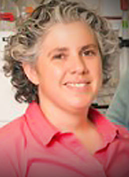
Melissa Farrow, PhD
Vanderbilt University
Dr. Farrow is a Research Associate Professor in Cell and Developmental Biology at University. She is the scientific director of the Biomolecular Multimodal Imaging Center and Associate Director of Research and Development for the Mass Spectrometry Research Center. Having served as the project manager across Hubmap, Kidney Precision Medicine Project, and Human Tumor Network Atlas consortia she has experience in the construction of molecular atlases across a variety of tissues in both two and three dimensional workflows.

Peter Sorger, PhD
Harvard Medical School
Peter Sorger is the Otto Krayer Professor of Systems Biology at Harvard Medical School, Head of the Harvard Program in Therapeutic Sciences and Director of the Laboratory of Systems Biology. His research focuses on single cell measurement and modeling of oncogenic networks in cancer and inflammatory diseases and on the interplay between tumor-intrinsic mechanisms of invasion and metastasis and countervailing immune surveillance. This is performed in human cohorts and mouse models to elucidate molecular programs controlling cancer progression and response to therapeutic drugs applied individually and in combination. His group is currently developing a range of experimental and computational methods for high-plex single-cell analysis of human tissue and tumors.

Robert Krueger, PhD
Department of Computer Science & Engineering at NYU Tandon
Robert Krueger is an assistant professor at New York University (NYU) - Department of Computer Science and Engineering, and a member of the NYU Visualization Imaging and Data Analysis Center. Previously, he was a postdoctoral research fellow and subgroup leader at VCG Harvard, School of Engineering and Applied Sciences at Harvard University, and a senior research scientist at the Laboratory of Systems Pharmacology, Harvard Medical School. Dr. Krueger received his Ph.D. degree (Dr. rer. nat.) in Computer Science at the Institute for Visualization and Interactive Systems, University of Stuttgart in 2017. Krueger's research interests lie in the field of data visualization and visual analytics for spatial and spatially-referenced multivariate data with a focus on biomedical visualization.

Sanjay Jain, MD, PhD
Department of Medicine - Division of Nephrology, Washington University School of Medicine
Sanjay Jain is a physician scientist focusing on basic translational research to understand fundamentals of cellular functions, organization and communication in the kidney and lower urinary tract in health and disease across life span. By building this foundational knowledge and multimodal and multiscalar 2D and 3D atlases, his team aims to glean insights into slowing disease progression and extending the life span of kidney function and hence the patient. This knowledge will be key in establishing the blueprint for engineering these organs to supplement kidney function. The Jain lab’s approach includes using model systems and human tissue based research integrated with clinicopathological data for discovery, validation and intervention in order to delineate mechanisms of kidney development, regeneration and repair. He has leading roles in several consortia including KPMP, HuBMAP and the Pediatric Centers of Excellence in Nephrology (PCEN).

Yuqi Tan, PhD
Stanford University
Dr. Tan is a computational biologist developing innovative tools to quantify cell identity, enhance stem cell engineering, and dissect cancer heterogeneity. During her Ph.D., she specialized in computational and quantitative analysis of single-cell RNA sequencing (scRNA-seq) data, contributing to multiple high-impact publications. As a postdoctoral researcher, she has advanced the integration of single-cell omics with multiplexed imaging to decode high-dimensional tissue architecture in cancer and psychiatric diseases. Her long-term vision is to leverage multi-omics and develop machine learning techniques for both 2D and 3D analysis to uncover how diverse cell types and their interactions shape development, aging, and disease.
Agenda
- 45 minutes
Breakfast
- 15 minutes
Welcome
- 1 hour
Keynote (Peter Sorger)
- 20 minutes
Coffee
- 1 hour 10 minutes
Talks (Yuqi Tan, Melissa Farrow, Robert Krueger)
- 1 hour 10 minutes
Talks (Matt Ruffalo, Gloria Pryhuber, Jakob Troidl)
- 1 hour 10 minutes
Lunch
- 1 hour 10 minutes
Talks (Clarence Yapp, Sanjay Jain, Eric Moerth)
- 1 hour
Keynote (Ashley Kiemen)
- 1 hour 30 minutes
Reception
bottom of page